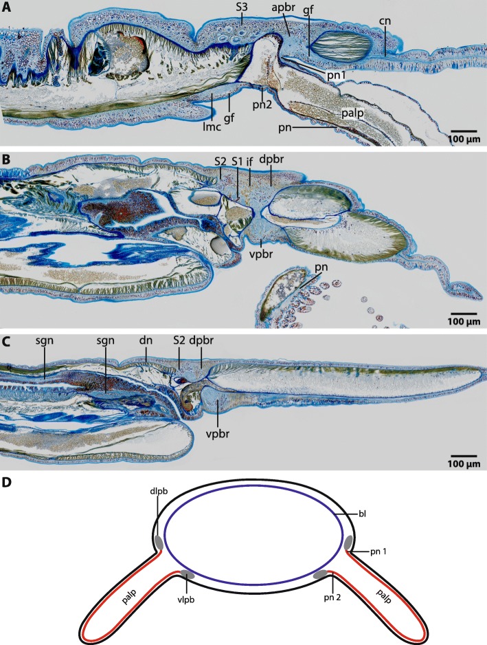
Fig. 4.
Magelona mirabilis, histological sagittal sections (5 μm), Azan staining a: The anterior part of the brain (apbr) is compact and gives rise to frontally located cephalic nerves (cn). A giant fibre (gf) originates in the ventral part of the brain and extends along the whole lengths of the lateral medullary cords (lmc). The palp nerve is connected to dorso- lateral and ventro- lateral part of the brain by two nerves (pn1, pn2). Neurons with very prominent somata (S3) are located posterior to the brain and are associated with the palp nerve. b: The dorsal part of the brain (dpbr) terminates in a layer of type two neurons (S2). A cluster of type one neurons (S1) is located between the neurites of the dorsal part of the brain. The palp is innervated by a basiepidermal palp nerve (pn). vpbr: ventral part of brain. Radial glia cells with prominent intermediate filaments (if) cross the neuropil. c: The dorsal part of the brain (dpbr) terminates in a layer of type two neurons (S2). A very delicate dorsal nerve originates in the neuropil of the posterior dorsal part of the brain (dpbr). The digestive system is innervated by stomatogastric nerves (sgn). vpbr: ventral part of brain. d: Palps are innervated by two nerves (pn1, pn2), of which one originates in the dorso- lateral (dlpb) and one in the ventro- lateral parts (vlpb) of the brain. bl: basal lamina

