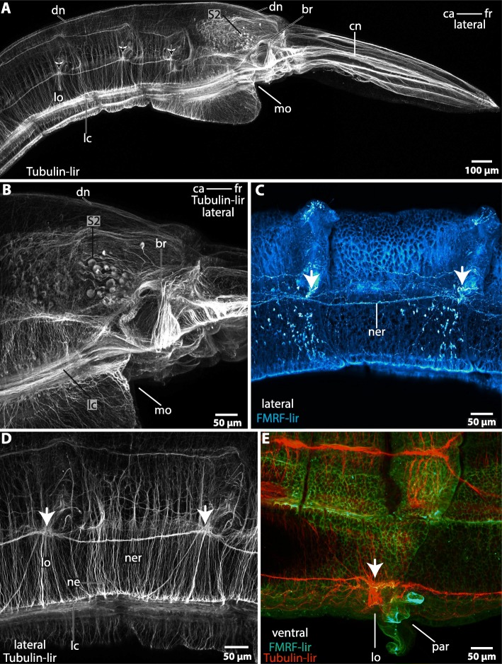
Fig. 6.
Magelona mirabilis, immunohistochemistry: a-d lateral view; e: ventral view. a: Tubulin-lir. A minor dorsal nerve (dn) arises in the posterior part of the brain (br). The tip of the head is innervated by cephalic nerves (cn) which originate in the brain (br). A lateral chain of neuropil concentrations (arrows) is connected to the brain. The concentrations correspond with the location of the lateral organ (lo). ca: caudal; fr: frontal; mo: mouth opening; S2: enlarged neurons. b: Tubulin-lir. In dorsal part of the brain (br) a cluster of enlarged neurons (S2) is present. The ventral part of the brain is confluent with the lateral medullary cords (lc). ca: caudal; fr: frontal; mo: mouth opening. c: FMRF-lir. Higher magnification of the lateral chain of neuropil concentrations in A. An accumulation of FMRF-positive neurites (arrow) is present at the heights of the lateral organ. The concentrations are interconnected by a nerve (ner). d: Tubulin-lir. Higher magnification of the lateral chain of neuropil knots in A. The ventro- lateral cords (lc) are connected to the chain of neuropil concentrations (arrows) by fine neurites (ne). e: Tubulin-lir, FMRF-lir, ventral view. The accumulations of neurites (arrow) are located close to the parapodia (pa) and correspond with the location of the lateral organ (lo). ca: caudal; fo: frontal

