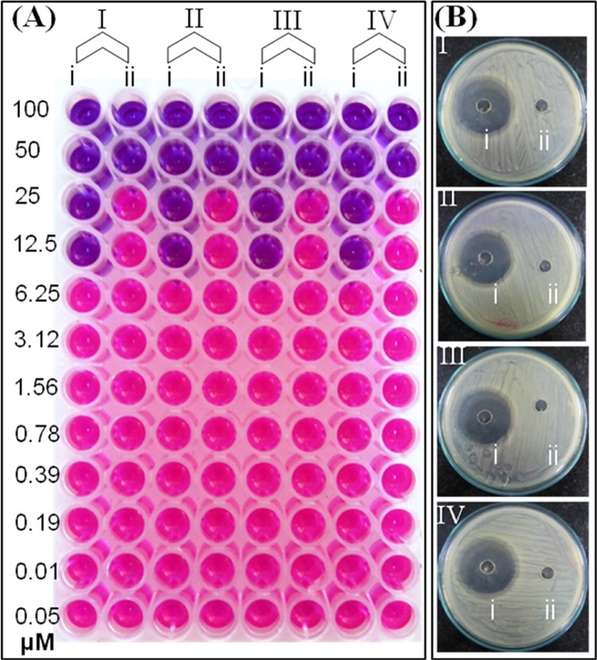
Figure 3.

Antibacterial activity of pCuS. (A) MIC was discerned from REMA. Blue color indicate dead cells, pink color denotes live cells, where (i)—pCuS and (ii)—BSA + pCuS. (B) Zone of inhibition assay was performed at 12.5 μM pCuS, where (i)—pCuS and (ii)—BSA + pCuS. I—E. coli, II—P. aeruginosa, III—B. subtilis, and IV—S. aureus. BSA—50 μM; jacalin—50 μM.
