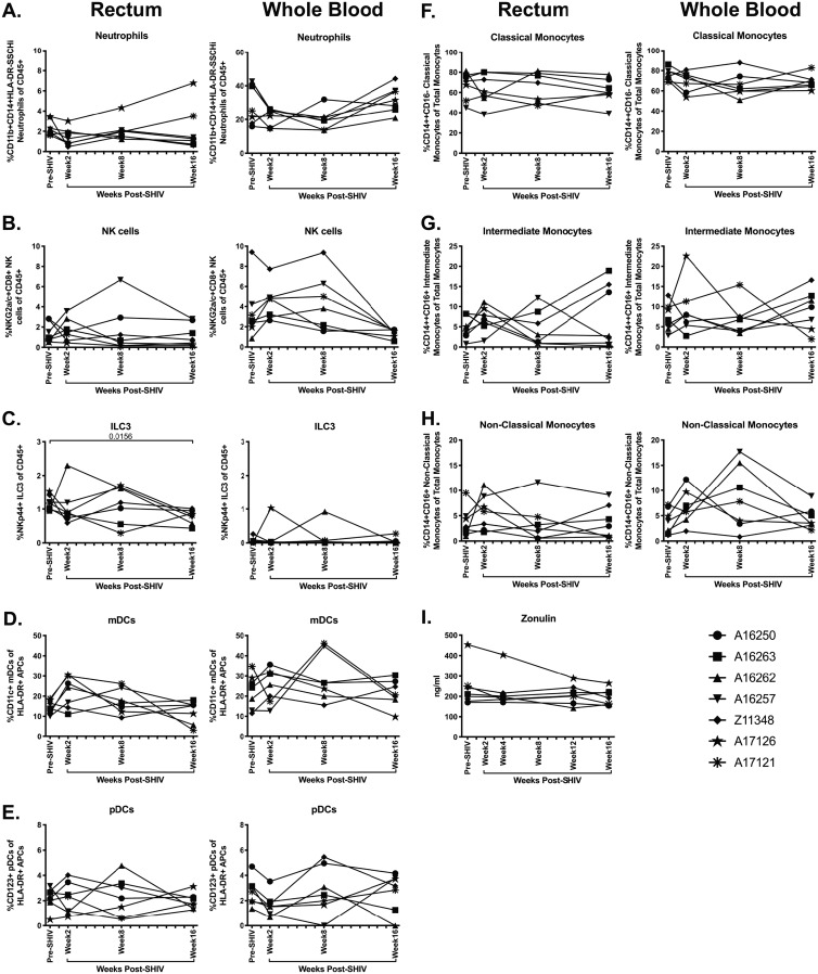
FIG 13.
Minimal alteration of innate immune subsets during acute SHIV.CH505 infection. Neutrophil, NK, ILC3, mDC, pDC, and monocyte subsets were characterized in rectum and whole blood of SHIV.CH505-infected rhesus macaques by flow cytometry. (A to C) Percentage of neutrophils (A) (CD11b+ CD14+ HLA-DR− SSChi), NK cells (B) (NKG2a/c+ CD8+), and ILC3s (C) (NKp44+ CD8−) of CD45+ cells in rectum and whole blood at all time points. (D and E) Percentage of mDCs (D) (CD11c+) and pDCs (E) (CD123+) of HLA-DR+ antigen-presenting cells in rectum and whole blood at all time points. (F to H) Percentage of classical monocytes (F) (CD14+ CD16−), intermediate monocytes (G) (CD14+ CD16+), and nonclassical monocytes (H) (CD14+ CD16+) of total monocytes in rectum and whole blood at all time points. (I) Plasma levels of zonulin-1 were quantified by ELISA. In all panels, each animal is represented by a different symbol. Statistical significance between post-SHIV infection time points and pre-SHIV infection baseline was calculated using Friedman’s test and Dunn’s multiple-comparison test.

