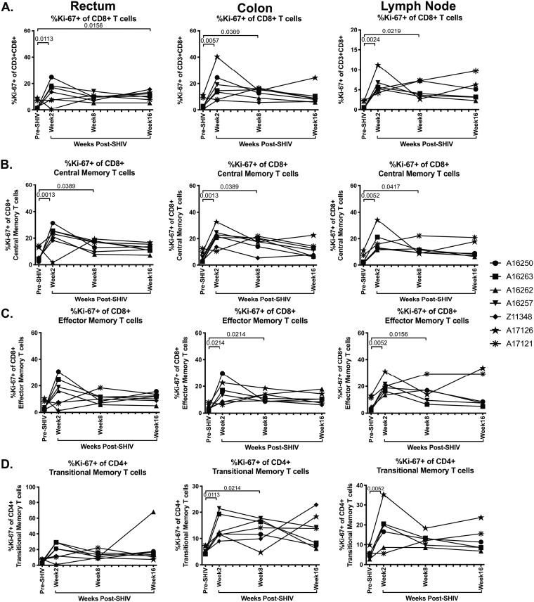
FIG 9.
Significant increase in Ki-67+ CD8+ T cells in SHIV.CH505 infection. CD8+ T cell subsets expressing Ki-67 were characterized in rectum, colon, and lymph node tissue of SHIV.CH505-infected rhesus macaques by flow cytometry. (A) Percentage of Ki-67+ cells of CD8+ T cells in rectum, colon, and lymph node at all time points. (B) Percentage of Ki-67+ cells of CD8+ central memory T cells in rectum, colon, and lymph node at all time points. (C) Percentage of Ki-67+ cells of CD8+ effector memory T cells in rectum, colon, and lymph node at all time points. (D) Percentage of Ki-67+ cells of CD8+ transitional memory T cells in rectum, colon, and lymph node at all time points. In all panels, each animal is represented by a different symbol. Statistical significance between post-SHIV infection time points and pre-SHIV infection baseline was calculated using Friedman’s test and Dunn’s multiple-comparison test.

