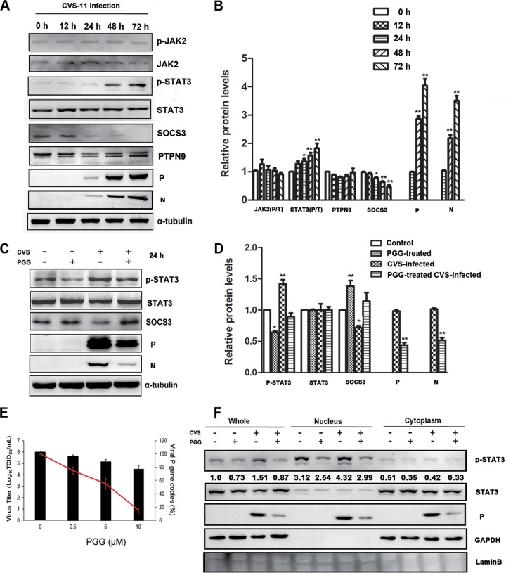
FIG 1.
RABV induced the activation of STAT3 via SOCS3, and PGG reversed it. BHK-21 cells were infected with CVS-11 for diverse times. (A) The expression levels of STAT3 pathway-associated proteins and the CVS P protein were detected by Western blotting. p-JAK2, phosphorylated JAK2. (B) The relative expression levels of the indicated proteins were analyzed by the use of ImageJ software and normalized to the α-tubulin expression level. BHK-21 cells were treated with CVS-11 or PGG alone or in combination. (C) The expression levels of SOCS3/STAT3 and the CVS P protein were detected by Western blotting. (D) The relative expression levels of the proteins were analyzed by the use of ImageJ software and normalized to the α-tubulin expression level. (E) The viral titer (bars) and P gene mRNA expression level (red line) of RABV were detected, respectively, by DFA and qRT-PCR. (F) BHK-21 cells were treated as described above for 48 h. Subcellular fractions of the cytosol (Cyt) and nucleus (Nuc) were isolated, and p-STAT3 and CVS P were analyzed by Western blotting. GAPDH and lamin B were used as purity controls for various fractions. The numbers indicate the ratio of p-STAT3/total STAT3. *, P < 0.05; **, P < 0.01. All data presented are representative of those from three independent experiments.

