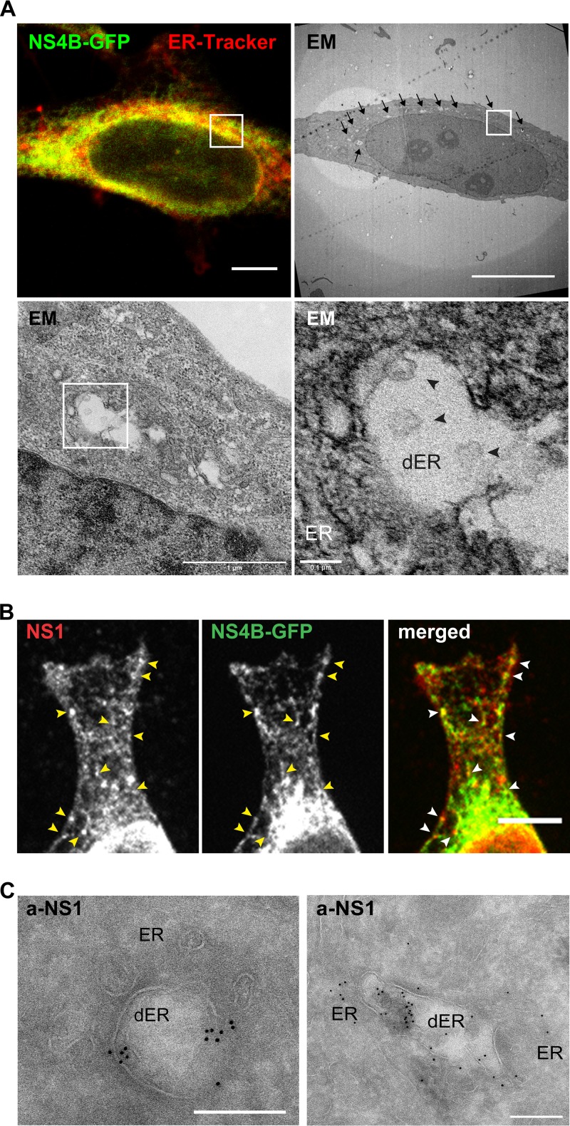
FIG 4.
Localization of NS proteins in RC-like structures in NSP-GFP cells. (A) CLEM images of a NSP-GFP cell induced with dox. (Top) The left fluorescence micrograph shows NS4B-GFP (green) and ER-tracker (red), and the right panel shows the correlated electron micrograph, with arrows highlighting dilated ER membranes. (Bottom) The left panel shows an electron micrograph of the area indicated by the white square in higher magnification, and the right panel shows further magnification of the indicated area; the ER and dilated ER (dER) are indicated, and black arrowheads denote vesicle-like structures. Scale bars = 10 μm (top) or as indicated below the bars (bottom). (B) Immunofluorescence micrographs of the single and merged channels of a dox-induced NSP-GFP cell expressing NS4B-GFP and stained against NS1. Yellow and white arrowheads exemplify the punctuate NS1 localization on NS4B-GFP-positive ER tubules. Scale bar = 10 μm. (C) Immuno-EM images of NSP-GFP cells induced with dox. The cells were stained with anti-NS1 antibodies conjugated with gold particles. Scale bars = 200 nm.

