FIG 5.
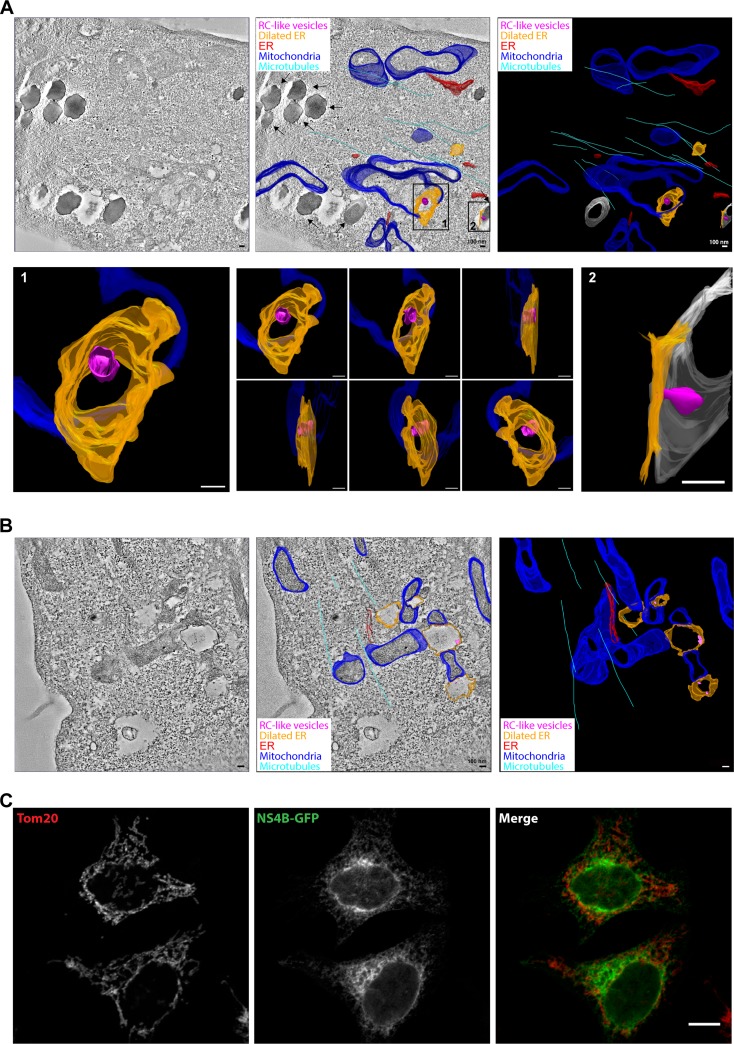
ER dilation caused by NS proteins is linked to mitochondria. (A and B) Electron tomography analysis of induced NSP-GFP cells, showing close contacts of lipid droplets, mitochondria, and dilated ER. Tomograms were recorded and the membranes of mitochondria, ER, lipid droplets, and microtubules were manually assigned, yielding a schematic, color-coded, 3D membrane model overlaid on representative tomograms. A tomographic slice (left), overlaid 3D model (middle), and 3D model (right) are presented to show dilated ER (orange) and RC-like invaginations (magenta). ER tubules are depicted in red, mitochondria in blue, microtubules in cyan, and lipid droplets in gray. Scale bars = 100 nm. (A) In the overlaid 3D model, black arrows indicate lipid droplets. Insets show magnification and different angles of the indicated areas (also see movie S2 in the supplemental material). (B) Representative electron tomograms of dox-induced NSP-GFP cells show close contact of mitochondria and dilated ER. (C) Fluorescence micrographs of NS4B-GFP in NSP-GFP cells costained for mitochondria using antibodies to the mitochondrial protein Tom20. Scale bar, 10 μm.

