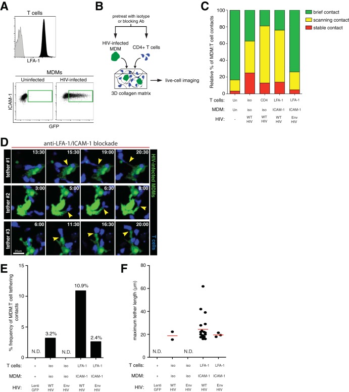
FIG 4.
Role of adhesive molecular interactions in stabilizing MDM-T cell contacts during HIV infection. (A) Cell surface LFA-1 expression on T cells (black) and isotype control staining (gray). ICAM expression on MDMs after infection with HIV-GFP for 2 days is boxed in green. (B) Schematic of the experimental setup. HIV-GFP-infected MDMs and CMTMR-labeled CD4+ T cells (blue) were pretreated with either isotype or the indicated blocking antibodies and embedded into a collagen matrix for live-cell imaging studies. (C) Relative proportions of MDM-T cell contacts characterized as stable, scanning, and brief. Blocking antibody conditions are indicated on the bottom axis. iso, isotype antibody. (D) Time series micrographs depicting three MDM-T cell-tethering events after LFA-1–ICAM-1 dual-antibody blockade. The arrowheads indicate the points of contact and the membranous tethers. The time stamps are in minutes. (E) Percent frequency of MDM-T cell-tethering events after specific antibody or isotype control blockade. Tethers were defined as membranous extensions between MDMs and T cells that were over 15 μm long. Frequency values (percent) are indicated above the bars. (F) Maximum tether lengths of MDM-T cell contacts. N.D., not detected.

