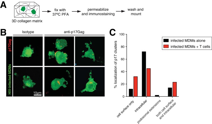
FIG 6.
Localization of VCCs in HIV-infected macrophages. (A) Schematic of the experimental setup. HIV-GFP-infected MDMs (green) were incubated for 24 h in collagen and then fixed with paraformaldehyde. Gels were prepared for immunostaining with anti-p17Gag antibody. (B) Confocal micrographs of HIV-GFP-infected MDMs (green) stained with either isotype or anti-p17Gag antibody (red). Clusters of p17Gag staining represent VCCs. (C) Percent localization of p17Gag+ VCCs in different regions of infected MDMs alone (n = 43) or after coculture with T cells (n = 65).

