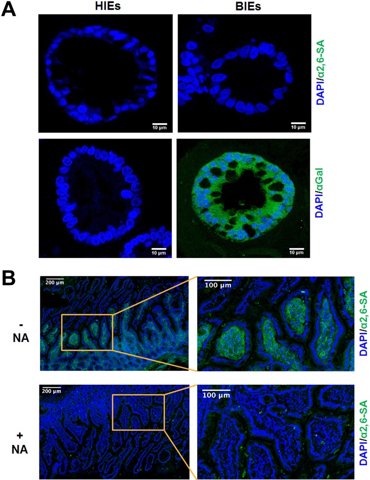
FIG 10.
Expression of α2,6-linked SA and αGal in differentiated human and bovine jejunal enteroids and human small intestinal sections. (A) Differentiated human jejunal and bovine ileal enteroids grown in Matrigel with a differentiation medium were fixed with 4% paraformaldehyde. Thin sections of differentiated HIEs and BIEs were stained using immunohistochemistry to determine the expression levels of α2,6-linked sialic acids and the αGal epitope. Nuclei were stained with DAPI. Representative images are shown. (B) Detection of α2,6-linked SAs in the human small intestine. Frozen human duodenal tissue sections were fixed with 10% formalin and incubated with biotin-labeled SNL at 10 μg/ml in PBS, followed by incubation with DyLight 488-conjugated streptavidin (green) in the same buffer (α2,6-SA). Nuclei were stained with DAPI (blue). To determine the SA dependence of the labeling, tissue sections were pretreated with neuraminidase for 4 h (+NA). Positive control sections were preincubated with the manufacturer’s enzyme buffer only (–NA). The right panel shows higher-magnification images of the inlets from the left panel. The data are representative from two individuals.

