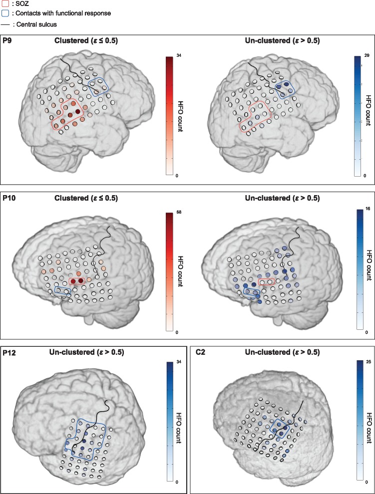
Figure 6.
Spatial projection of repetitive and non-repetitive HFOs in four patients. In each patient the individual MRI together with the 3D electrode model generated directly from the co-registered CT image is shown. The ‘repetitive’ events are defined as the HFOs being clustered within a radius of 0.5. The results are shown for Patients 9 and 10 where both SOZ and functional cortex were covered by the grid electrodes, and for Patient 12 and Control 1 where the electrodes included the motor cortex only. Compared to the HFOs identified by using a ε ≤ 0.5, the remaining unclustered HFOs are spatially localized at functional cortex or brain regions distinct from the SOZ.

