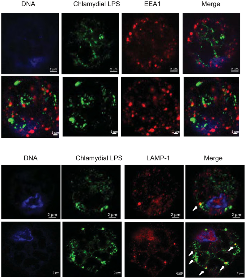
Fig 3: At 2 h post-infection, both C. muridarum and C. caviae do not co-localize with early endosomes, but more numbers of C. caviae co-localize with lysosomes.
Macrophages infected with C. muridarum or C. cavaie at 1 MOI infection for 3 h p.i were fixed and immuno-stained for an early endosomal marker EEA-1 (A) or lysosomal marker LAMP-1 (B) with chlamydial LPS staining. Arrows indicate co-localization of LAMP-1 and LPS. Image shows a single cell for clarity. Larger field is shown in Fig S3. A representative image from one of two experiments is shown.

