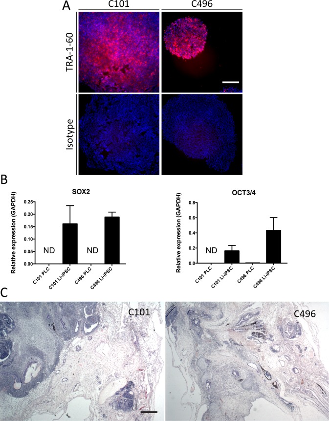
Fig 2. Pluripotency of Li-iPSCs.
(A) Surface pluripotency marker TRA-1-60 specific or isotype IgG life immunofluorescence analysis of Li-iPSC colonies derived from patients C101 and C496 (scale bar 250μm). (B) Pluripotency gene (SOX2 and OCT3/4) expression in PLCs and Li-iPSCs derived from patients C101 and C496 was quantified by RT-qPCR using total cellular RNA. Shown is the relative expression of the transcripts compared to GAPDH (mean +/- SD; ND = not detectable). (C) Hematoxylin and eosin staining of formalin fixed and paraffin embedded sections from teratomas isolated from NOD-SCID mice 50 days after subcutaneous injection of Li-iPSCs from patients C101 or C496 (scale bar 500μm).

