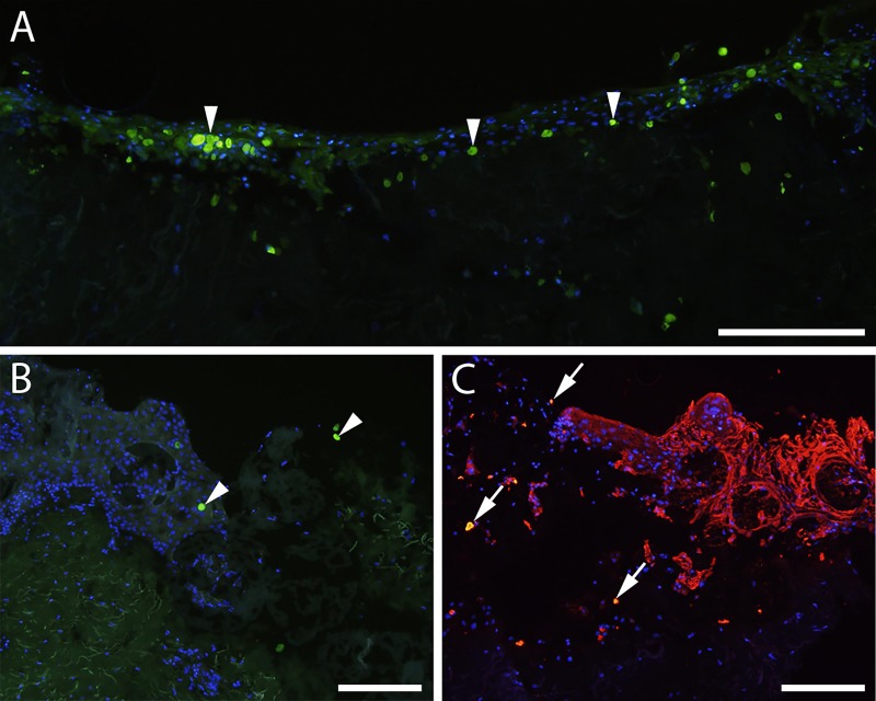
Fig 4. Transplantation of CFSE-stained keratinocytes to ex vivo wounds.
(A) 5(6) carboxyfluorescein-N-hydroxysuccinimidyl ester (CFSE)-stained keratinocytes are incorporated into the neoepidermis in a fully re-epithelialized ex vivo wound seven days post transplantation, seen as green stained cells. (B) CFSE-stained keratinocytes (green) on microcarriers in an ex vivo wound seven days post transplantation. Arrowheads indicate CFSE-stained keratinocytes. (C) CFSE-stained keratinocytes on microcarriers in an ex vivo wound 21 days post transplantation. Double staining with CFSE and antibodies against cytokeratin detected with red fluorescent secondary antibodies can be identified by the yellow color (arrows), confirming that the stained cells are transplanted keratinocytes. Scale bars = 200 μm.

