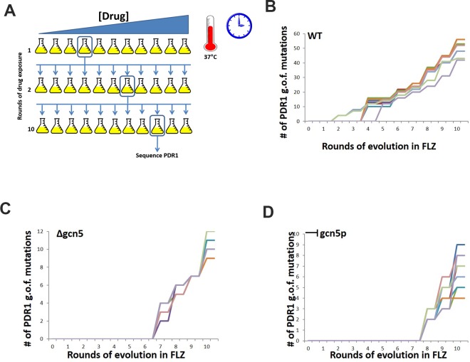
Fig 5. Evolution of wildtype, gcn5 null and gcn5 chemically inhibited C. glabrata cells in the presence of fluconazole.
C. glabrata cells grown in increasing concentrations of FLZ, with the PDR1 gene sequenced after each round to determine when and in which domain gain of function mutations are identified in. (A) Schematic of experiment. 10 individual flasks of C. glabrata cells – wildtype, Δgcn5 and chemically inhibited Gcn5 were exposed to increased concentrations of FLZ. The flask where inhibition of growth was first observed was used as the started culture for the subsequence round of drug exposure until 10 rounds of drug exposure was completed. At each pitching of cells, PDR1 was sequenced to identified when a gain of function mutation first emerged. (B) In wildtype C. glarbata cells, after 3 rounds of exposure to increasing FLZ concentrations, PDR1 mutations were identified and mapped to the activation domain. (C) C. glabrata Δgcn5 cells, after 6 rounds of exposure to increasing FLZ concentrations, PDR1 gain of function mutations were isolated and mapped to the activation domain and the middle homology domain. (D) C. glabrata cells that had Gcn5p chemically inhibited through with the addition of ɣ-, after 7 rounds of exposure to increasing FLZ concentrations, the emergence of PDR1 gain of function mutations was observed in the activation domain and DNA binding domain. The number of gain of function mutations observed is dramatically reduced in both the Δgcn5 and chemically inhibited Gcn5 FLZ exposures.

