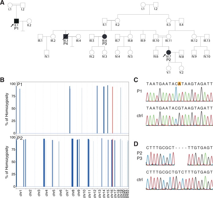
Fig 1. Pedigrees and genetic findings.
(A) Pedigrees of the patients analyzed in this study. DNA was available only for subjects P1, P2, and P3. (B) Exome-wide homozygosity mapping for autosomal chromosomes, using the AutoMap tool. The autozygous region containing the gene ARL2BP is highlighted in red. (C and D) Sanger validation of the WES findings, showing the presence of a homozygous splice site mutation in patient P1 (C, NM_012106.3:c.207+1G>A, leading to p.Asp35PhefsTer8), and a frameshift deletion in patients P2 and P3 (D, NM_012106.3:c.33_36delGTCT: p.Phe13ProfsTer15), alongside with relevant control sequences (ctrl).

