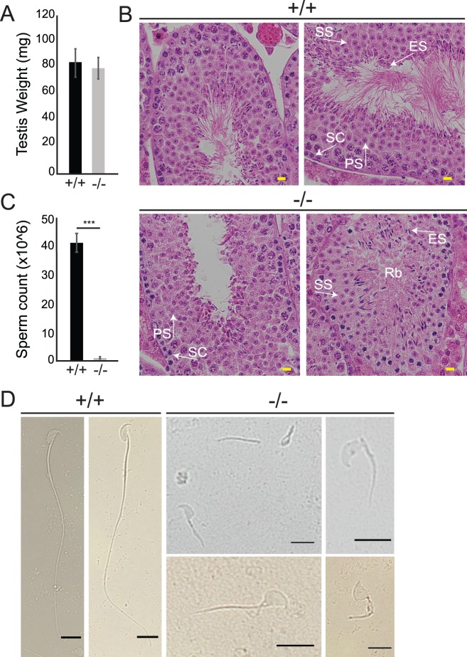
Fig 5. Loss of ARL2BP leads to decreased sperm count and abnormal sperm structure in mice.
(A) The average weight of testis between WT (+/+) and KO (-/-) mice are comparable, according to unpaired, two-tailed t test (n = 10). Data are represented as the mean ± SEM. (B) H&E sections of WT (+/+) and KO (-/-) testis. Residual bodies (Rb), PS = primary spermatocyte, SS = secondary spermatocyte, ES = elongating spermatid, SC = sertoli cells. Scale Bar = 20μm. (C) Graph presenting the epididymal sperm cell counts of WT (+/+) and KO (-/-) mice. Data are represented as the mean ± SEM. ***P = 0.0002, according to unpaired, two-tailed t test (n = 3). (D) Light microscopy images displaying WT (+/+) sperm with normal sperm structure, while KO (-/-) sperm show abnormal sperm heads, shorter sperm tails, detached head and tails, and retained cytoplasm. Scale Bar = 10μm.

