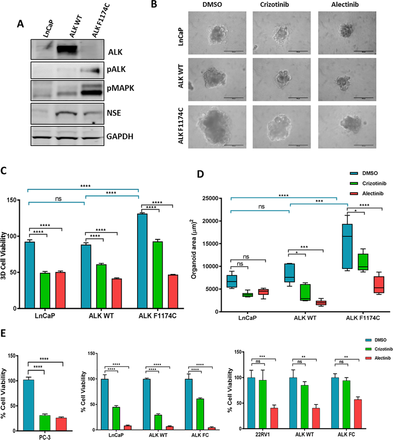
Figure 3. Alectinib suppresses ALK F1174C induced proliferation in prostate cancer cells.
(A) Western blot analysis of ALK, pALK, pMAPK and NSE in LNCaP cell infected with ALK WT and ALK F1174C expressing lentivirus. GAPDH and Tubulin are internal loading controls. Data represent three independent experiments. (B) Representative images showing growth of LNCaP organoids Cells were infected with ALK WT or ALK F1174C and grown in matrigel supplemented media to form organoids for 7 days. At day 8, organoids were treated with 5μm crizotinib or alectinib for 7 days. Scale bars: 100μm. (C) Quantification of percent 3D cell viability assay (D) Quantification of organoid area (μm2). (E) Percent cell viability after 72 h treatment with 10μm crizotinib or alectinib in PC-3, LNCaP and 22Rv1 cells. Data represent mean ± standard error, n=6. ns=non significant, *P ≤ 0.05, **P ≤ 0.01, ***P ≤ 0.001, ***P ≤ 0.001.

