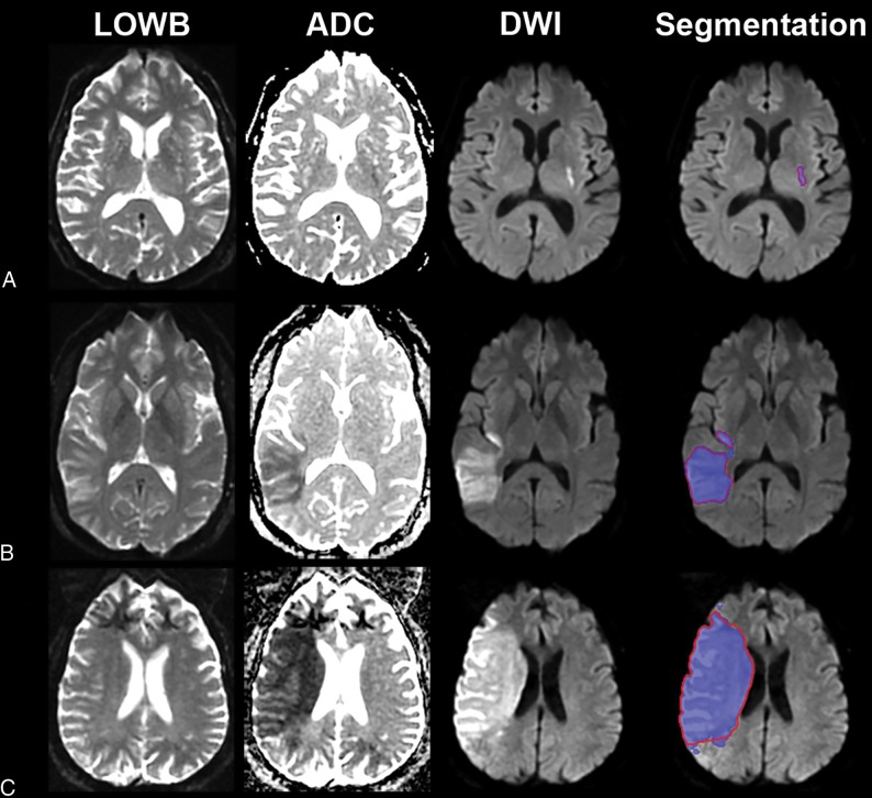
Fig 2.
Sample segmentation results of the ensemble of DWI+ADC+LOWB (blue regions) on sample subjects along with manual outlines (red outlines). A, A small lesion example from a 70-year-old man with an admission NIHSS score of 1, imaged approximately 9 hours from LKW: MLV = 0.96 cm3, ALV = 1.07 cm3, Dice = 89.4%. B, Medium lesion sample from a 38-year-old woman with an admission NIHSS score of 4, imaged approximately 10 hours from LKW: MLV = 54.3 cm3, ALV = 57.9 cm3, Dice = 95.7%. C, A large lesion example from a 62-year-old man with an undocumented admission NIHSS score, imaged approximately 10 hours from LKW: MLV = 229.0 cm3, ALV = 208.7 cm3, Dice = 94.0%.

