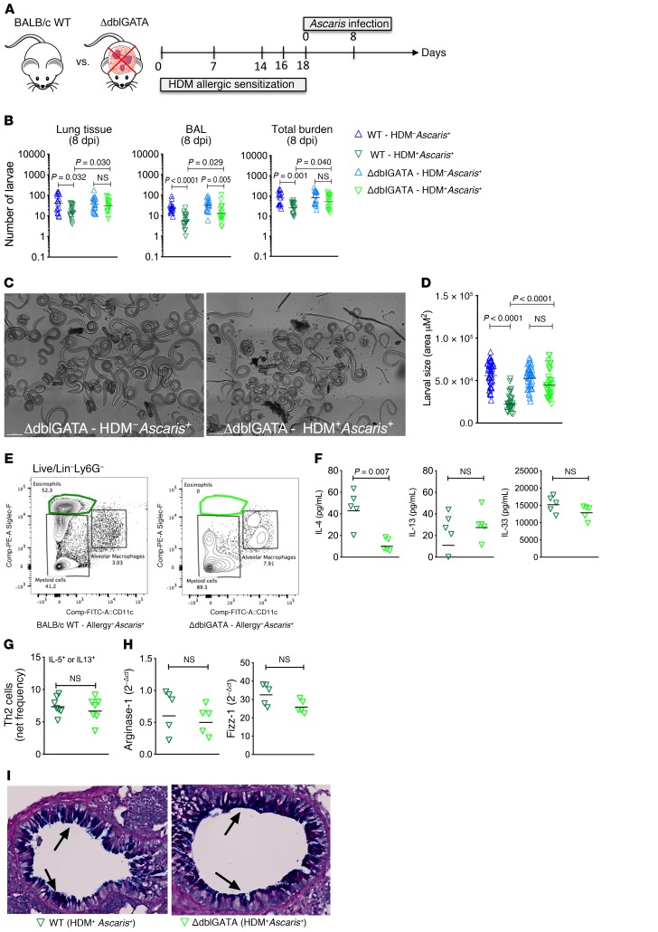
Figure 5. Ascaris larval migration and development occur normally in allergic eosinophil-deficient mice.
(A) Experimental design scheme for HDM allergic sensitization followed by Ascaris infection in WT BALB/c and eosinophil-knockout mice (ΔdblGATA). (B–D) Total parasite burden in the lungs and BAL at day 8 of infection (n = 17 mice per group) (B), as well as a representative bright confocal image of lung-stage larvae recovered from Ascaris-infected ΔdblGATA mice following or not following HDM allergic sensitization (HDM–Ascaris+ vs. HDM+Ascaris+) (scale bars: 200 μm) (C) and a morphometric analysis of these larvae recovered (D). Three independent experiments were performed. (E–I) Characterization of the lung-specific immune response of HDM+Ascaris+ WT (n = 5 mice) (dark green triangles) and HDM+Ascaris+ ΔdblGATA mice (n = 5 mice) (light green triangles) by flow cytometry phenotypic analysis, cytokine production, M2 macrophage marker gene expression, and histology of lung epithelium. (E) Eosinophils. (F) IL-4, IL-13, and IL-33 cytokine quantification. (G) IL-5– or IL-13–producing CD4+ Th2 cell frequency (n = 7 mice per group). (H) Arginase-1 and Fizz-1 gene expression in the lungs. (I) Lung sections stained by AB/PAS showing mucus production by the goblet cells in the lung epithelium (in blue; arrows) (original magnification, ×16). Each symbol represents a single mouse, and the horizontal bars are the GMs. P values are indicated in each graph. Differences between HDM+Ascaris+ (WT) and HDM+Ascaris+ (GATA) mice were considered statistically significant at P < 0.05 by unpaired Mann-Whitney test. *Significantly different (P < 0.05) from naive (HDM–Ascaris–) group. Kruskal-Wallis test followed by Dunn’s multiple-comparisons test was used to compare WT HDM+Ascaris+ versus ΔdblGATA HDM+Ascaris+ in B and D.

