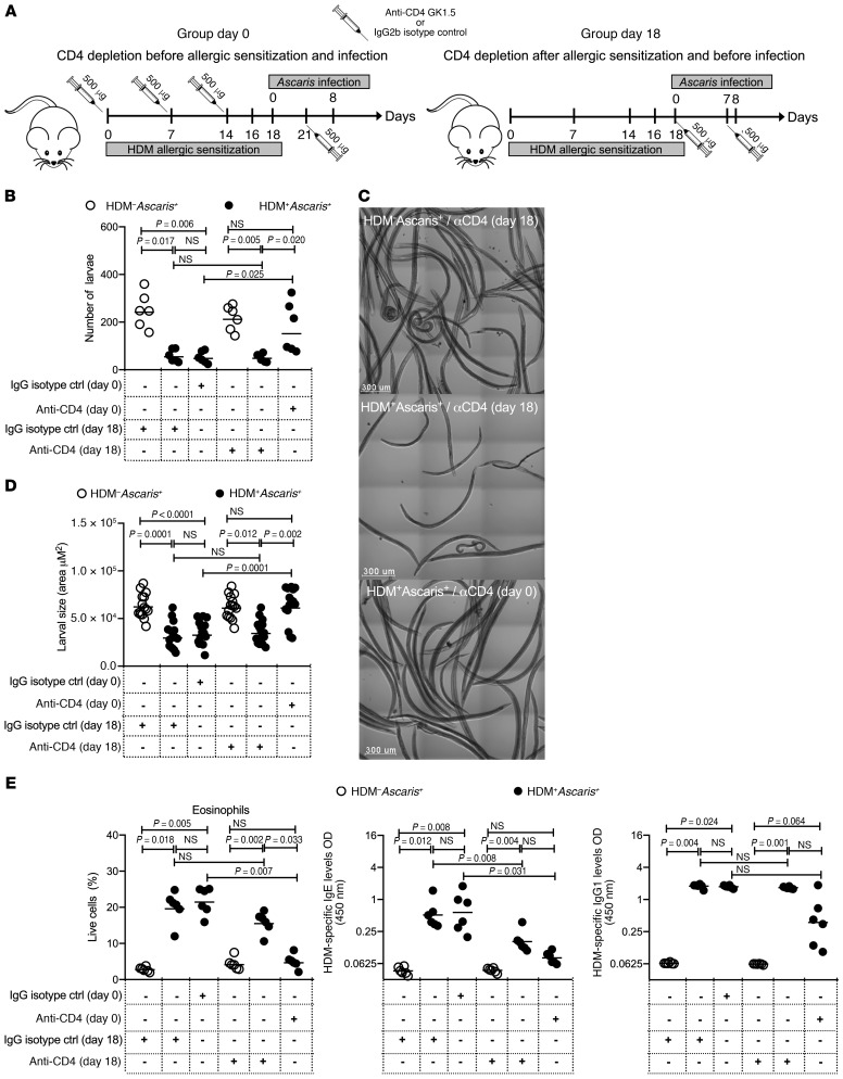
Figure 6. HDM-induced immunity to Ascaris parasites is dependent on eosinophils driven by CD4+ T cells.
(A) Experimental design scheme for CD4+ T cell depletion in unsensitized mice followed by Ascaris infection: HDM–Ascaris+ (open circles in B, D, and E) and HDM-sensitized Ascaris-infected (filled circles in B, D, and E) mice received anti-CD4 antibodies before and continuously during the HDM sensitization at days 0, 7, 14, and 21 (group day 0), or after allergic sensitization at days 18 and 25 (group day 18) (n = 6 mice per group). Rat IgG2b antibody was used as the isotype control. (B) Total parasite burden in the BAL at 8 dpi. (C) Representative bright confocal images of the larvae recovered in the BAL at 8 dpi (scale bars: 300 μm). (D) Lung-stage larval development by morphometric analysis of larva size recovered from HDM–Ascaris+ and HDM+Ascaris+ treated with anti-CD4 or isotype control. (E) Characterization of the lung-specific immune response by flow cytometry, showing the frequency of Siglec-F+CD11c– eosinophils and serum HDM-specific IgE and IgG1 antibody levels. Each symbol represents a single mouse, and the horizontal bars are the GMs. P values are indicated in each graph. Nonparametric Kruskal-Wallis test followed by Dunn’s multiple-comparisons test was used for differences among the isotype control–treated groups and anti-CD4–treated groups, and the differences for HDM+Ascaris+ animals treated with IgG2b or anti-CD4 antibodies were considered statistically significant at P < 0.05 by Kruskal-Wallis test followed by Dunn’s multiple-comparisons test.

