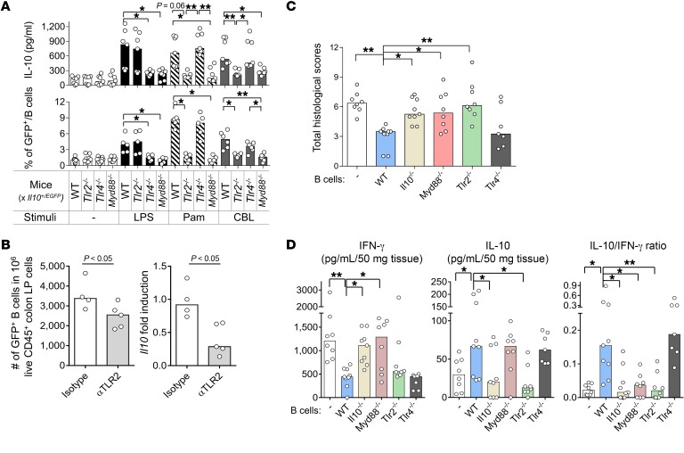
Figure 7. TLR2/MyD88 signaling increases the frequency of IL-10–producing B cells upon bacterial product stimulation ex vivo and mediates the suppression of experimental colitis by IL-10–producing B cells in vivo.
(A) Colonic LP B cells from 8- to 10-week-old WT Il10+/EGFP, Tlr2−/− Il10+/EGFP, Tlr4−/− Il10+/EGFP, and Myd88−/− Il10+/EGFP mice were cultured without (–) or with 10 μg/mL CBL, 50 ng/mL Pam3, or 200 ng/mL LPS for 24 hours, after which IL-10 levels in culture supernatants were measured by ELISA (top panel). GFP+ populations in B cells (live CD45+CD19+B220+) were analyzed with flow cytometry in reference to WT (GFP–) control cells stained with the same antibodies as target samples. Data are presented as median of 7–8 separate cell cultures, with cells in each culture pooled from 2–3 mice, combined from 3 independent experiments (bottom panel). *P < 0.05, **P < 0.01. (B) Eight-week-old GF Il10+/EGFP reporter mice were conventionalized with SPF fecal bacterial transplantation. Mice were given anti-TLR2 antibody (αTLR2) or isotype control i.v. on days 0 and 4 after fecal bacterial transplantation and harvested on day 7 for IL-10 analysis. Colonic LP cells were isolated, and live CD45+GFP+ B cells (CD19+B220+) were analyzed by flow cytometry (left) as described above; Il10 mRNA levels were normalized with Actb (right). n = 4–5 mice. Data are presented as median, Mann-Whitney U test. (C and D) Splenic B cells (1 × 10⁶) from SPF-raised WT, Tlr2−/−, Tlr4−/−, Myd88−/−, or Il10−/− mice were cotransferred with 5 × 10⁵ WT naive CD4+ T cells isolated by a naive CD4+ T cell isolation kit (see Methods, Cell purification) into SPF Rag2−/− Il10−/− recipients. Six weeks after cell transfer, mice were evaluated for (C) severity of colitis by histology and (D) measurement of spontaneously secreted IL-10 and IFN-γ in colonic tissue explants by ELISA. n = 7–9 mice/group, combined from 2 independent experiments. Data are presented as median; *P < 0.05 and **P < 0.01 (vs. WT mice), Kruskal-Wallis test with Dunn’s post hoc test.

