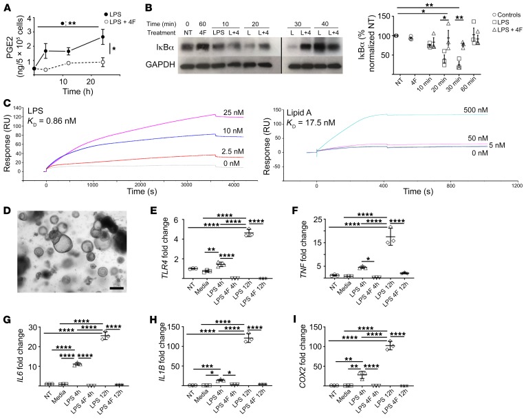
Figure 6. 4F inhibits the LPS-mediated proinflammatory response of human macrophages and intestinal epithelium.
(*P < 0.05; **P < 0.01; ***P < 0.001; ****P < 0.0001. (A–B) Human THP1 macrophages were treated with 4F (15 μg/mL), LPS (20 ng/mL), or LPS + 4F. (A) 4F significantly inhibited total PGE2 in cell lysates and media for 24 hours (n = 3/group). (B) 4F (4) significantly inhibited LPS dependent (L) IκBα degradation at 30 minutes, as determined by Western blot (NT, no treatment) (left: representative blot; right: densitometric analysis of 3 experiments) (noncontiguous sections of same blots). (C) 4F binds both LPS (left; KD = 0.86 nM) and lipid A (right; KD = 17.5 nM) with high affinity, as determined by surface plasmon resonance analysis. (D) Crypts were isolated from human small intestine and grown into enteroids in matrigel (representative image; scale bar: 200 μm). (E–I) Conditioned media from THP-1 cells treated with LPS or LPS + 4F for 4 hours (LPS 4h; LPS + 4F 4h) or 12 hours (LPS 12h; LPS + 4F 12h) was added to the enteroids, and proinflammatory gene expression at 12 hours was determined by qPCR (n = 3/group; Media indicates THP1 media only; fold change vs. NT). For statistical analyses, 1-way (B, E) or 2-way (A) ANOVA with Tukey’s multiple comparisons test and adjusted P values were used.

