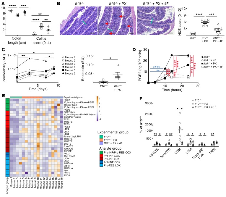
Figure 9. 4F treatment inhibits the development of colitis in the piroxicam-accelerated Il10–/– model of IBD.
*P < 0.05; **P < 0.01; ***P < 0.001; ****P < 0.0001. (A–B) Il10–/– mice were treated with and without piroxicam (PX) and 4F (n = 4–7/group). (A) 4F partially rescued the effect of PX on colon length and colitis score. (B) 4F also improved disease histopathology: representative H&E images (scale bars, 200 μm) and H&E disease score are shown. Green bars indicate thickness of mucosa in Il10–/– + PX. Red arrows indicate inflammatory foci. (C) PX increased both whole intestinal barrier permeability of Il10–/– mice in a biphasic manner, as determined by LC-MS/MS of urinary excretion of sucralose (left), as well as the translocation of endotoxin into portal vein serum at day 9 (right). (D) 4F inhibited the production of prostanoids including PGE2 in LPS-activated Il10–/– BMDMs with or without PX (n = 3/group). (E–F) The lipid inflammatory mediator profile in the colons of the mice shown in A and B was determined by LC-MS/MS (see Supplemental Table 8) (n = 4–8 mice/group). (E) Heatmap of changes. (F) All significant differences are represented. 4F significantly inhibited the disease-dependent increase of proinflammatory LOX mediators. Tt, total. Statistical analyses were as follows: 1-way (A, B), repeated measures 1-way (C, left), or 3-way (D) ANOVA with Tukey’s multiple comparisons test and adjusted P values; Student’s t test (C, right). For E–F, we used the Benjamini-Hochberg procedure applied to 1-way ANOVA for each lipidomic analyte with FDR at level α = 0.05, followed by Tukey’s multiple comparisons test and adjusted P values.

