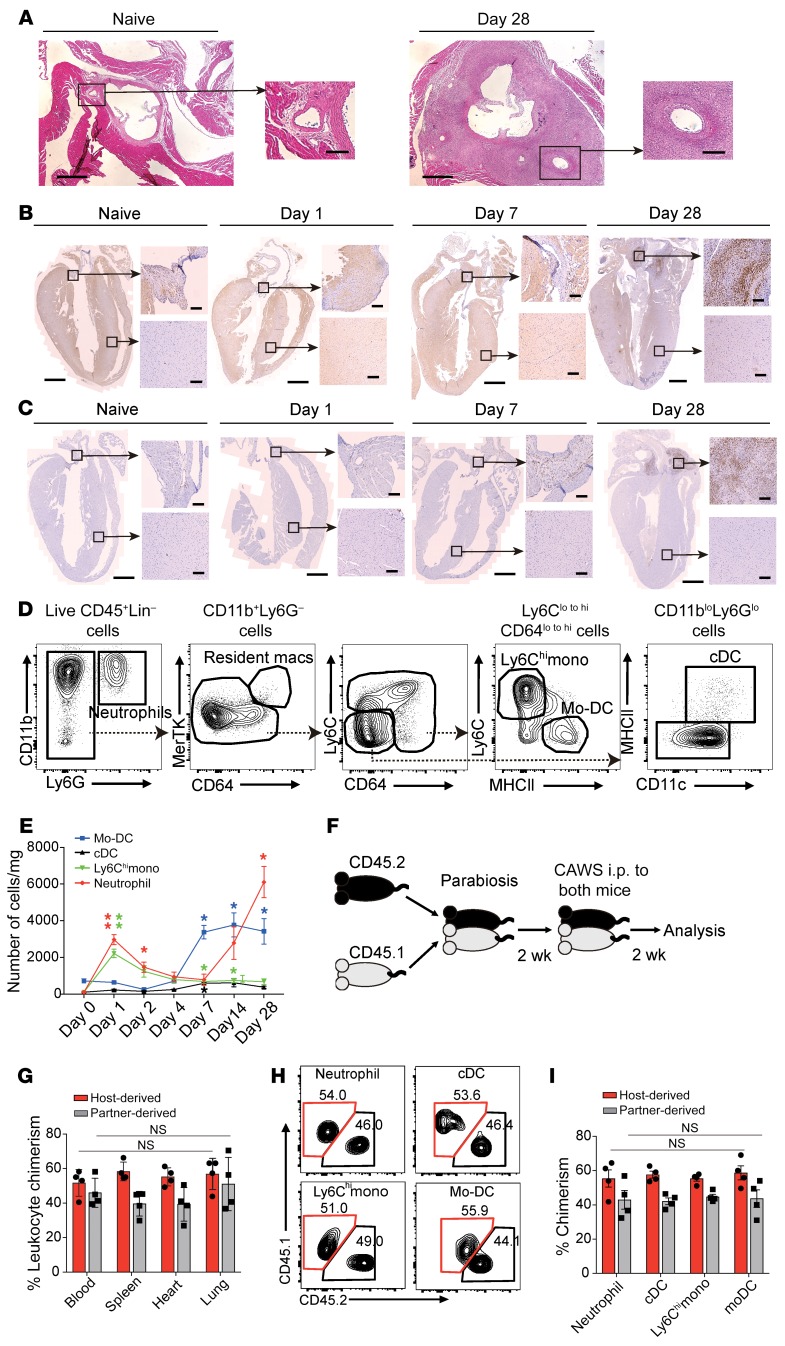
Figure 1. CAWS induces inflammatory monocyte recruitment into the heart on day 1.
(A) Representative H&E–stained horizontal section of the aortic root area from WT naive mice or on day 28 after 5 daily i.p. injections of CAWS beginning on day 0. Low-power field shows aortic root area and high-power field shows the ostium of the coronary artery within the aortic root. Scale bars: 400 μm (low-power images) and 100 μm (high-power images). (B and C) Coronal section isolated from naive or CAWS-injected WT mice (on day 1, 7, or 28) stained with anti-Ly6G/Ly6C (B) or anti-F4/80 (C) for IHC. Cropped high-power-field images show aortic root (upper panel) or myocardium (lower panel). Scale bars: 1 mm (low-power images) and 100 μm (high-power images). (D) Representative FACS analysis of cardiac neutrophil, macrophage, DC, and monocyte subsets recovered from CAWS-injected mice on day 1 after the first CAWS injection. One representative of 3 independent experiments. (E) Kinetics of absolute cell numbers of the indicated immune cell subset per mg of heart isolated during the course of CAWS-induced vasculitis (mean ± SEM, n = 4–5 mice per time point, *P < 0.05, **P < 0.01 versus day 0, using unpaired 2-tailed Student’s t test). (F) Schematic protocol for parabiosis experiment. (G) Tissue chimerism was analyzed in parabiotic pairs by measuring the frequency of host-derived leukocytes (CD45.1+) and partner-derived leukocytes (CD45.2+) on day 14 after CAWS injection (mean ± SEM, n = 4 mice). NS indicates statistically identical in the indicated organs among host-derived cells or partner-derived cells using ordinary 1-way ANOVA with Tukey’s post hoc test. (H) Representative FACS plots of each myeloid cell subset in the parabiotic mouse hearts on day 14 after the first CAWS injection. (I) Quantitation of chimerism for the indicated myeloid cell subsets (mean ± SEM, n = 4 mice). All values were statistically identical in indicated cell subsets among host-derived cells or partner-derived cells using ordinary 1-way ANOVA with Tukey’s post hoc test. cDC, cardiac DC.

