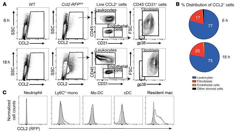
Figure 3. Cardiac-resident macrophages are the main source of early CCL2 production.
(A) Representative contour plots of RFP+ CCL2-expressing cells in live singlets derived from WT or Ccl2-RFPfl/fl reporter mouse hearts 6 hours and 18 hours after CAWS injection. One representative of 3 independent experiments. (B) Pie chart showing percentage distribution of RFP+ cells for the indicated subpopulations in Ccl2-RFPfl/fl reporter mice heart 6 hours and 8 hours after CAWS injection. (C) Representative histograms of CCL2 expression in myeloid populations isolated from the hearts of Ccl2-RFPfl/fl reporter mice 1 day after CAWS injection (n = 5 mice). Individual leukocyte populations were immunophenotyped based on the gating strategy shown in Figure 1A. The RFP reporter was used to identify CCL2-producing cells.

