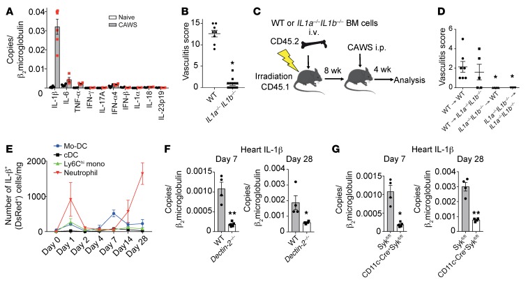
Figure 7. Dectin-2–dependent production of IL-1β from CD11c+ cells is required for CAWS-induced vasculitis.
(A) Heart tissue from WT mice was harvested 28 days after initial CAWS injection and assessed for cytokine expression by qPCR (n = 4–5 per group, mean ± SEM). (B) Vasculitis scores of IL1a–/– IL1b–/– mice or WT mice 28 days after CAWS injection (mean ± SEM, *P < 0.0001 versus WT). (C) Schematic of BMC mice generation using WT and IL1a–/– IL1b–/– mice. (D) Vasculitis scores were assessed 28 days after CAWS injection into 8-week-reconstituted WT → WT, WT → IL1–/– IL1b–/–, IL1a–/– IL1b–/– → IL1a–/– IL1b–/–, and IL1a–/– IL1b–/– → WT BMC mice (mean ± SEM, *P < 0.01 versus WT → WT). (E) Kinetics of IL-1β+ (DsRed+) cell numbers of the indicated immune cell subset per mg of heart tissue determined by flow cytometric analysis on day 0, 1, 2, 4, 7, 14, or 28 after CAWS injection of pIl1-DsRed mice (mean ± SEM, n = 3 per time point). (F) Dectin-2–/– or WT mice were injected with CAWS and 7 or 28 day later hearts were harvested and assessed for IL-1β expression by qPCR (mean ± SEM, n = 4–5 per group). (G) CD11cΔSyk mice or control mice were injected with CAWS and 7 or 28 day later hearts were harvested and assessed for IL-1β expression by qPCR (F and G; mean ± SEM, *P < 0.05, **P < 0.001 versus Sykfl/fl). P values were calculated using unpaired 2-tailed Student’s t test (B, F, and G) or 1-way ANOVA with Dunnett’s post hoc test (D).

