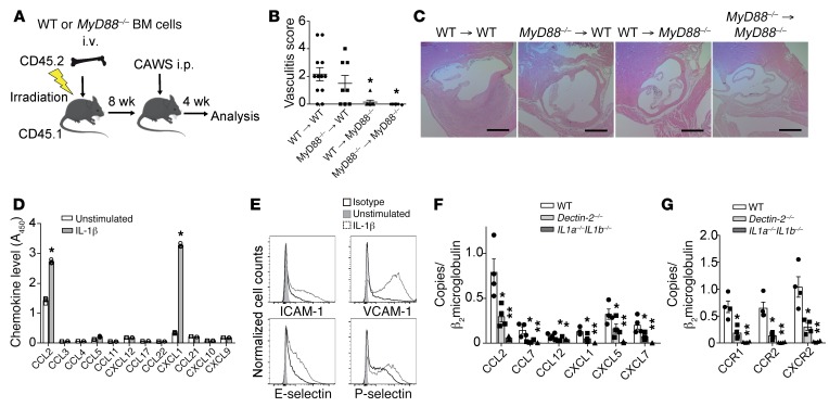
Figure 9. IL-1β/MyD88 signaling in heart stromal cells is required for chemokine and adhesion molecule induction and the development of vasculitis.
(A) Schematic of BMC mice generation using WT and MyD88–/– mice. (B) Vasculitis scores were assessed 28 days after CAWS injection into 8-week-reconstituted WT → WT or WT → MyD88–/– versus MyD88–/– → MyD88–/– or MyD88–/– → WT BMC mice (mean ± SEM, n = 7–12 mice per group, *P < 0.01 versus WT). (C) H&E staining of aortic root lesions isolated from the indicated BMC mice on day 28. Scale bars: 400 μm. (D and E) Mouse aortic endothelial cells were stimulated with IL-1β (10 ng/mL) for 18 hours and chemokine protein levels in the culture supernatant were measured by ELISA (mean ± SEM, n = 3, *P < 0.0001 versus unstimulated) (D). The expression levels of adhesion molecules were assessed by flow cytometry (E). One representative of 3 independent experiments. (F and G) qPCR analysis for chemokines (F) and chemokine receptors (G) in heart tissues isolated from WT, Dectin-2–/–, or IL1a–/– IL1b–/– mice 28 days after CAWS injection (mean ± SEM, n = 4–6 mice per group, *P < 0.05 versus WT, **P < 0.01 versus WT). P values were calculated using unpaired 2-tailed Student’s t test (D) or 1-way ANOVA with Dunnett’s post hoc test (B, F, and G).

