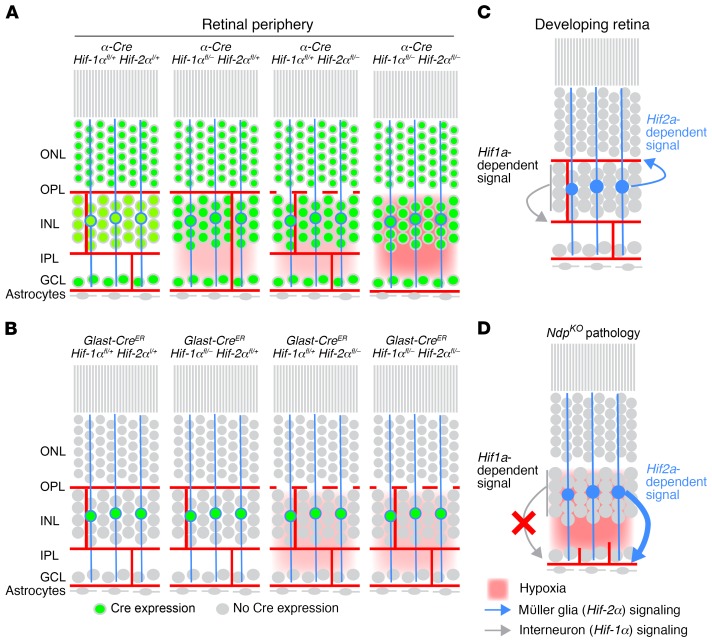
Figure 2. Complementary roles for Hif-1α and Hif-2α in the development of intraretinal capillaries.
(A and B) The principal cell layers of the adult retina, with neuronal and astrocyte cell bodies (grey), Müller glia cell bodies and processes (blue circles and vertical lines), retinal vasculature (red lines), extent of hypoxia (red background), and presence of Cre-mediated recombination (green circles; representing Cre activation of a nuclear localized GFP reporter). Summary phenotypes are shown for α-Cre (A) or Glast-CreER (B) mediated deletion of the indicated Hif-1α and/or Hif-2α alleles. (C and D) Retinal schematics as in A and B showing the cellular sources, vascular targets, and relative strengths of Hif-1α–dependent and Hif-2α–dependent proangiogenic signals during the final phase of retinal development (C) and in response to hypoxia in the mature NdpKO retina (D).

