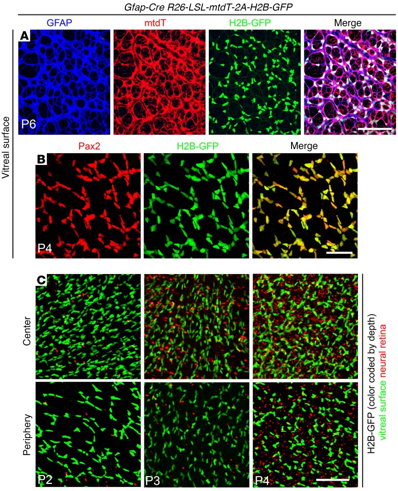
Figure 4. Highly efficient Cre-mediated recombination in early postnatal retinal astrocytes by the Messing Gfap-Cre line, as seen in retina flat mounts from Gfap-Cre R26-LSL-mtdT-2A-H2B-GFP retinas between P2 and P6.
(A and B) Retinas imaged at the vitreal surface and showing GFAP, mtdT, and H2B-GFP at P6 (A) and Pax2 and H2B-GFP at P4 (B). (C) H2B-GFP at the vitreal surface (green) and in the neural retina (red) at P2, P3, and P4. Highly efficient recombination in surface astroctyes is followed by scattered recombination in cells that are deeper within the neural retina. See Figure 7A for a cross-sectional view. Scale bars: 100 μm (A), 50 μm (B), and 100 μm (C).

