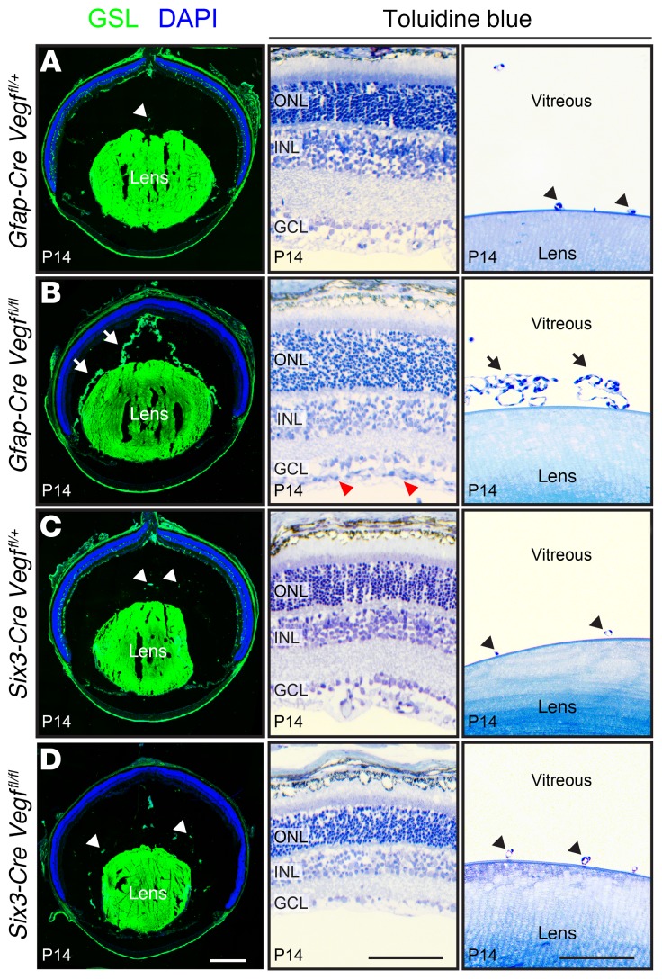
Figure 6. Persistent hyaloid vasculature in eyes without astrocyte-derived VEGF.
(A–D) The left column shows frozen eye sections stained with GSL at P14. The hyaloid vasculature is retained in Gfap-Cre Vegffl/fl eyes (arrows in left panel of B) and regresses in Gfap-Cre Vegffl/+, Six3-Cre Vegffl/+, and Six3-Cre Vegffl/fl eyes (arrowheads in left panels of A, C, and D). The right 2 columns show semithin plastic sections stained with toluidine blue (middle and far-right panels of A–D). In Gfap-Cre Vegffl/fl eyes, there is hyperproliferation of astrocytes on the vitreal face of the retina (red arrowheads in middle panel of B) and retention of hyaloid vessels near the lens (retrolental vessels; arrows in far-right panel of B). In the 3 other genotypes, the retrolental vasculature has almost completely regressed (arrowheads in far-right panels of A, C, and D). Representative frozen sections are shown from experiments with a total of 4 eyes per genotype, and representative plastic sections are shown from experiments with a total of 2 eyes per genotype. Scale bars: 500 μm (left panels of A–D) and 100 μm (middle and far-right panels of A–D).

