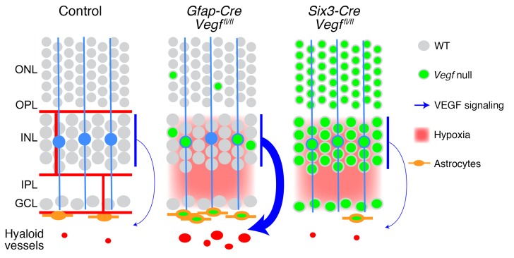
Figure 8. Retinal diagram as in Figure 2A showing the cellular sources, vascular targets, and relative strengths of VEGF signals inferred from the phenotypes of WT, Gfap-Cre Vegffl/fl, and Six3-Cre Vegffl/fl retinas.
The persistent hyaloid vasculature in Gfap-Cre Vegffl/fl retinas is presumed to reflect high levels of retina-derived VEGF.

