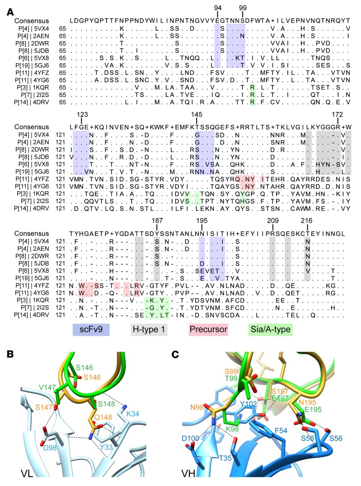
Figure 7. Structural basis of the genotypic specificity of human mAb9.
(A) Structure-based sequence alignment of VP8*s. The scFv9 and the known glycan binding sites in VP8*s are denoted in different colors: the scFv9 binding residues are shown in blue, the H-type 1 HBGA binding site in gray, the precursor HBGA binding residues in red, and the A-type HBGA-interacting residues in P[14] and the sialic acid binding residues in P[3] and P[7] in green. The PDB ID is given for each structure. (B and C) Superposition of scFv9/P[4] VP8* structure with that of P[6] RV3 VP8* (PDB ID: 5VX8) showing how the sequence changes in P[6] VP8* abrogates its binding to scFv9. The P[4] and P[6] VP8*s are colored in green and yellow, respectively; the VL and VH of scFv9 are shown in light and dark blue, respectively.

