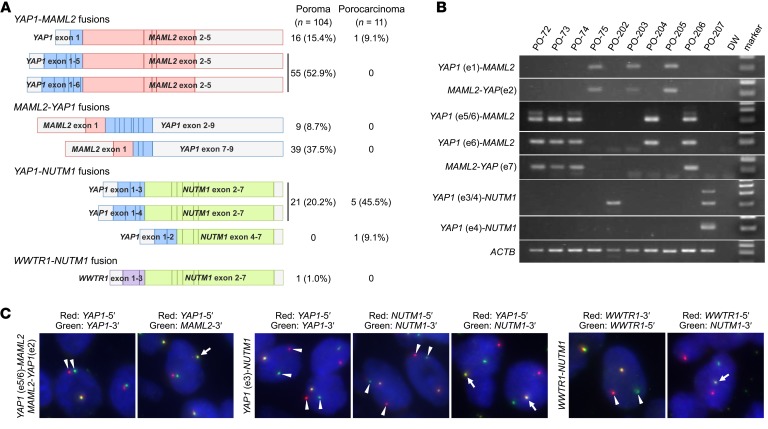
Figure 1. Detection of YAP1 and WWTR1 fusions in poromas and porocarcinomas.
(A) Fusion gene transcripts detected in poromas and porocarcinomas. All of the MAML2-YAP1 fusions were associated with the reciprocal YAP1-MAML2 fusions. (B) Representative gel images of RT-PCR products. MAML2-YAP1 fusion transcripts were consistently associated with the reciprocal YAP1-MAML2 fusions. Many of the tumors expressed multiple transcriptional variants. ACTB served as a positive control. (C) Representative fluorescence in situ hybridization images. The detected fusion transcripts are indicated on the left of the respective images. Arrowheads, split signals. Arrows, fused signals.

