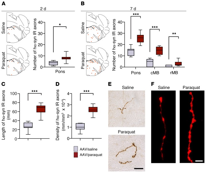
Figure 5. Oxidative stress promotes caudo-rostral spreading of hα-synuclein in the mouse brain.
Mice received an infusion of hα-synuclein–carrying AAVs into the left vagus nerve and were injected i.p. with either saline or paraquat and sacrificed at 2 or 7 days after treatment. Tissue sections were immunostained with anti–hα-synuclein. (A and B) The schematic plots show the distribution of hα-synuclein–labeled axons (each red dot represents one of these axons) in the left (AAV-injected side) pons. In the graphs, data show the counts of hα-synuclein–immunoreactive axons in the left pons at 2 and 7 days and in the caudal (cMB) and rostral midbrain (rMB) at 7 days. Tissue was obtained from mice treated with hα-synuclein AAVs/saline (n = 6 at 2 days and n = 7 at 7 days, gray bars) or with hα-synuclein AAVs/paraquat (n = 7 at 2 days and n = 9 at 7 days, red bars). (C and D) Length and density of hα-synuclein–positive axons were estimated at 7 days after treatment in a pontine area encompassing the locus coeruleus and the nucleus parabrachialis using the Spaceballs stereological tool. Analyses were made in the left pons in samples collected from mice treated with hα-synuclein AAVs/saline (n = 7) or with hα-synuclein AAVs/paraquat (n = 9). (E and F) Representative images of pontine axons immunolabeled with anti–hα-synuclein and visualized using brightfield (brown) or fluorescent (red) microscopy. Scale bars: 20 μm (E); 5 μm (F). Box and whisker plots show median, upper and lower quartiles, and maximum and minimum as whiskers. *P ≤ 0.05; **P ≤ 0.01; ***P ≤ 0.001, Mann-Whitney U test.

