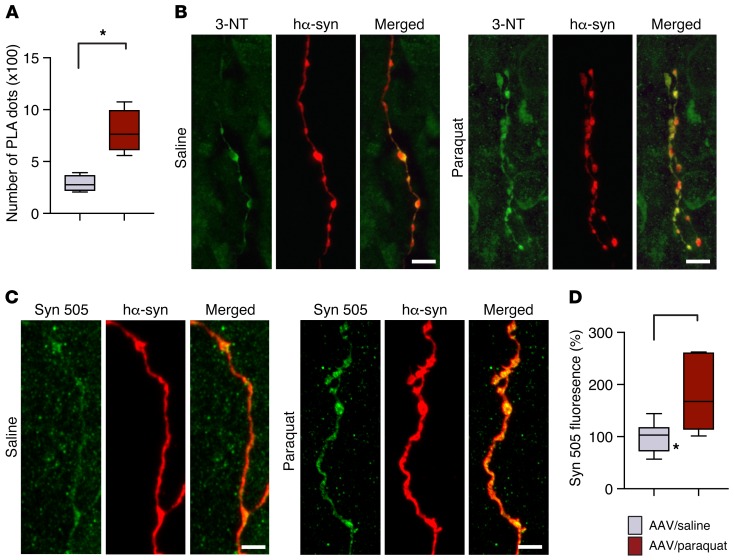
Figure 6. Oxidatively modified hα-synuclein is accumulated within pontine axons during caudo-rostral spreading of the protein.
Mice received an infusion of hα-synuclein–carrying AAVs into the left vagus nerve and were injected systemically with either saline or paraquat and sacrificed at 7 days after treatment. (A) Nitrated hα-synuclein was detected by indirect hα-synuclein/3-NT PLA. The number of PLA dots in the left pons from mice treated with hα-synuclein AAVs/saline (n = 4, gray bar) or with hα-synuclein AAVs/paraquat (n = 5, red bar) was counted. (B) Pontine tissue sections were costained with anti–3-NT and anti–hα-synuclein. Representative confocal images show labeled axons in the left pons. Scale bars: 5 μm. (C) Representative confocal images show axons in the left pons stained with anti-Syn 505 and anti–hα-synuclein. Scale bar: 5 μm. (D) Measurements of Syn 505 fluorescence were carried out in the left pons of mice treated with hα-synuclein AAVs/saline (n = 7) or with hα-synuclein AAVs/paraquat (n = 6). At least 3 axons/animal were analyzed and averaged. Values are expressed as percentage of the mean value in hα-synuclein AAV/saline-injected animals. Box and whisker plots show median, upper and lower quartiles, and maximum and minimum as whiskers. *P ≤ 0.05, Mann-Whitney U test.

