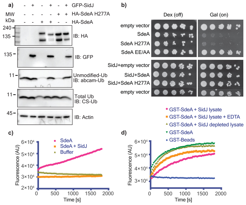
Figure 1. SidJ inhibits the Ub-ADP-ribosylation activity of SdeA.
a) SidJ and SdeA constructs were expressed as indicated in HEK293T cells and ubiquitin modification was probed using the Ub antibodies abcam-Ub and CS-Ub as described previously3. b) Yeast strain W303 was transformed using the indicated combination of constructs. Serial dilutions of transformed yeast were spotted on dextrose (repressing) or galactose (inducing) containing plates. SdeA EE/AA indicates mutation of E860, E862 to alanine. c) Purified SdeA from HEK293T cells expressing SdeA alone or in combination with SidJ was used in ε-NAD+ hydrolysis assays. Increase in the fluorescence indicates Ub-ADP ribosylation20. d) SdeA purified from E.coli was incubated with HEK293T cell lysate containing SidJ or depleted of SidJ. SdeA was subsequently purified using glutathione agarose beads and used in ε-NAD+ hydrolysis assays. Experiments in panels a-d were repeated thrice independently with similar results. For gel source data, see Supplementary Fig. 1.

