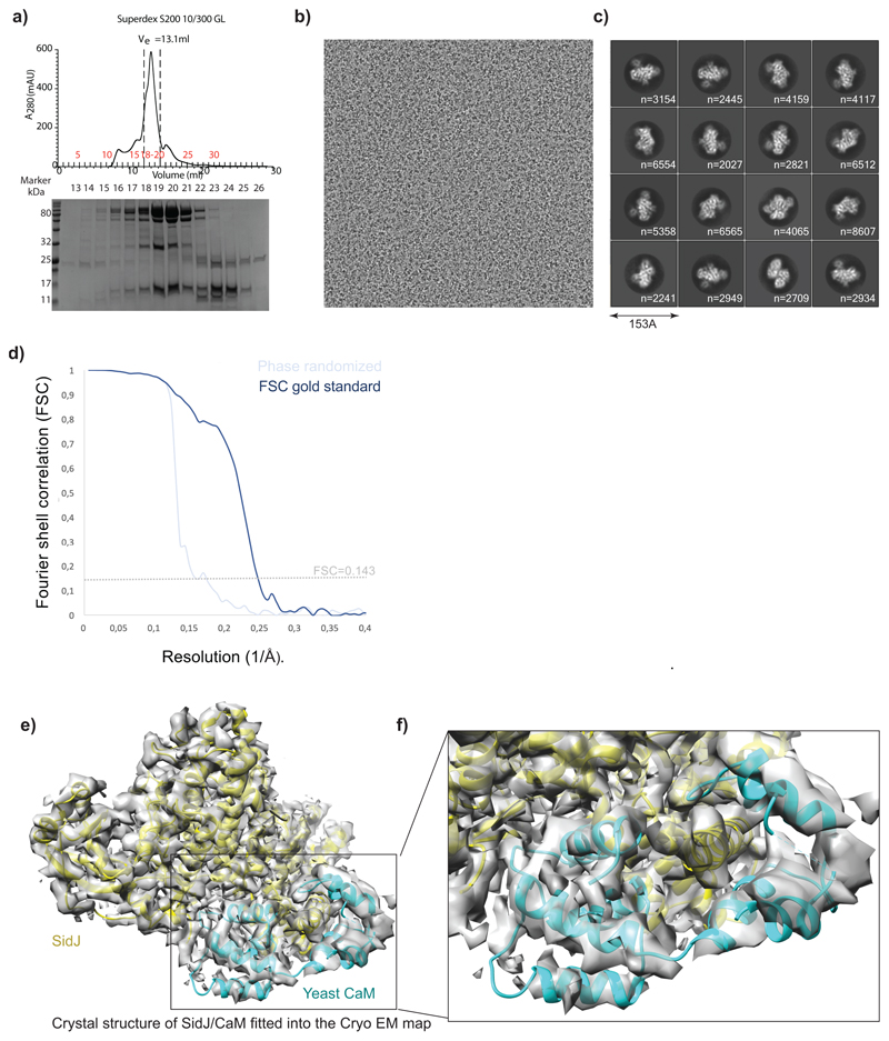
Extended data Fig. 6. Cryo-EM data processing and 3D reconstruction.
a) Size-exclusion profile of SidJ/CaM complex, elution fractions were analysed by SDS PAGE. Marked fractions were used for cryo-EM sample preparation. This experiment was repeated thrice independently with similar results. b) A representative electron micrograph for the cryo-EM dataset collected. c) Reference-free representative 2D class averages of the SidJ/CaM complex. Secondary structure features are already visible in projection images. Used number of particles to obtain a 2D class average is mentioned accordingly in each subpanel.
d) Gold-standard Fourier Shell Correlation21 plot between two independently refined half-maps, FSC0.143=4.15 Å resolution. FSC between phase-randomized half-maps show, as expected, a rapid drop of correlation beyond randomization point. e) Crystal structure of SidJ/CaM (PDB:6OQQ) is fitted into the cryo-EM 3D reconstruction f) A part of panel d is magnified to highlight the difference between the crystal structure and the cryo-EM map.

