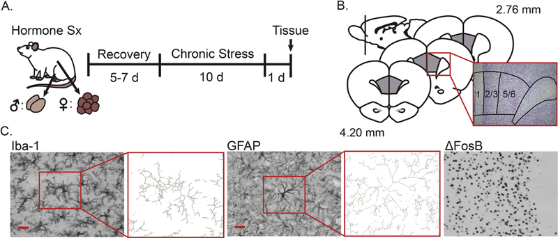
Fig 1.

Experimental design and histological analyses in prelimbic cortex. A. Male and female rats underwent either sham surgery or hormone manipulation (male: castration with or without hormone replacement, female: ovariectomy with or without hormone replacement), after which they were given 5–7 days to recover. Rats were then restrained for 3 hours/day over 10 days, or were left unhandled except for weighing every other day. Approximately 24 h after the final stressor, rats were euthanized and brains were collected, sectioned, and stained. Sx: Surgery. B. Prelimbic cortex was identified based on cytoarchitecture. C. Microglia (Iba-1) and astrocytes (GFAP) were visualized, and neuronal activation was inferred from ΔFosB expressing cells. Microglial and astrocyte morphology were characterized using a standardized threshold technique (immunopositive area) and skeleton analyses (process complexity = z-normalized number of branch points + z-normalized branch length / 2). Scale bar = 25 μm.
