Fig 7.

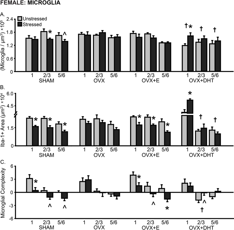
Layer-specific effects of gonadal hormones and stress on microglial density and morphology in prelimbic cortex in female rats. A. Total microglial density based on estimated volume. The density of microglia was reduced in OVX+DHT compared to SHAM females. Stress reduced microglial density in SHAM females yet increased this in OVX+DHT animals. B. Total area of Iba-1+ material. Stress reduced the area of microglial material in SHAM and OVX+E females across all layers, whereas treatment with DHT led to a stress-induced increase in Iba-1+ area in layer 1 in OVX females. C. Process complexity per microglia. There was decreased microglial complexity in layer 2/3 in OVX+DHT females. Stress induced a tendency toward- or significantly- decreased microglial complexity in SHAM and OVX+E females. OVX: Gonadectomized females without hormone treatment. +E: Treated with estradiol. +DHT: Treated with dihydrotestosterone. †p < .05 compared to same layer SHAM group. *p < .05, ^ p < .10 compared to same hormone condition unstressed group. X-axis labels represent layer (1, 2/3, 5/6). Error bars indicate SEM. Number of animals per group: SHAM female (unstressed: 7–8, stressed: 8–9), OVX (unstressed: 7, stressed: 7–8), OVX+E (unstressed: 7–8, stressed: 7–8), OVX+DHT (unstressed: 8, stressed: 7). SHAM outlier removed from microglial density (unstressed: 1, n = 1; stressed: 2/3, n = 1) and area (unstressed: all layers, n = 1; stressed: 1, n =1) analyses. OVX+E outlier removed from microglial density (unstressed: 2/3, n = 1) and area (unstressed: 1, n = 1; stressed: 2/3, n =1) analyses.
