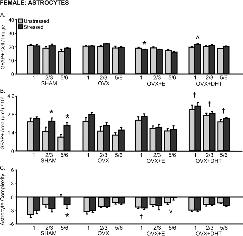
Fig 8.

Layer-specific effects of gonadal hormones and stress on astrocyte counts and morphology in prelimbic cortex in female rats. A. Number of GFAP+ cells counted per image analyzed. B. Total area of GFAP+ material. Treatment with DHT increased GFAP+ area in OVX females. Stress increased the area of astrocyte coverage in layers 2/3 and 5/6 in SHAM females. C. Process complexity per astrocyte. Stress reduced astrocytic complexity in layer 5/6 in SHAM females. OVX: Gonadectomized females without hormone treatment. +E: Treated with estradiol. +DHT: Treated with dihydrotestosterone. †p < .05, v p < .10 compared to same layer SHAM group. *p < .05, ^ p < .10 compared to same hormone condition unstressed group. X-axis labels represent layer (1, 2/3, 5/6). Error bars indicate SEM. Number of animals per group: SHAM female (unstressed: 7–8, stressed: 7–8), OVX (unstressed: 7, stressed: 8), OVX+E (unstressed: 8, stressed: 8), OVX+DHT (unstressed: 8, stressed: 7). SHAM outlier removed from astrocyte area analysis (unstressed: 5/6, n = 1; stressed: 1, n = 1).
