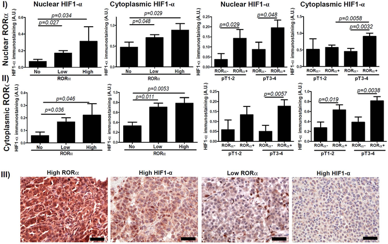
Figure 2.
Relationship between HIF-1α and RORα immunostaining. Differences in nuclear (I) and cytoplasmic (II) HIF-1α immunostaining in relation to nuclear (upper panel) and cytoplasmic (middle panel) RORα in primary melanomas. Statistically significant differences are denoted with p values as determined by Student’s t-test. III) Representative immunostaining of RORα and HIF-1α in melanoma patient with high RORα and high HIF-1α and in melanoma patient with low RORγ and low HIF-1α. Scale bars - 50μm (lower panel).

