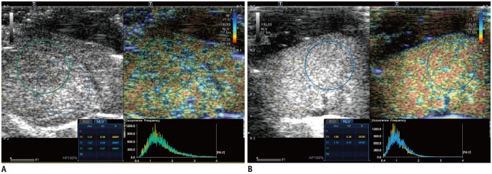
Fig. 4. Representative examples of NLV examinations in rat livers without (A) and with (B) hepatic steatosis.
A. Three ROIs were placed in right lobe of liver in rat from control group. Obtained median NLV value was 1.52, and histopathologic examination revealed no steatosis (not shown). B. Two ROIs were drawn in both lobes of liver in rat fed MCD diet for 2 weeks. Obtained median NLV value was 1.04, and histopathologic examination demonstrated severe steatosis (> 66%) (not shown).

