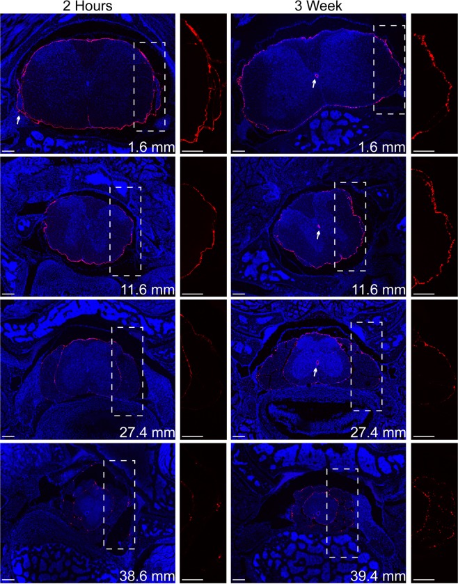
Figure 2.
Representative confocal images of FNP distribution along the spinal cord at 2 hours and 3 weeks. FNPs (red) distributed to the sacral regions of the spinal column within 2 hours and remained for over 3 weeks. FNPs could be observed following the meninges around spinal nerve roots (2 hr arrow), which may represent a clearance route for FNPs from the CNS. Even at later time points, the FNPs were mainly confined to meninges with minimal evidence for parenchymal penetration. FNPs appeared more punctate at later time points and could frequently be found within the central canal (3 wk arrows). The location of the slice along the spinal column is shown for each slice in the lower right. Zoomed in image of only FNP signal within the dashed box shown to the right of each tissue slice. Equivalent linear adjustments were made to the FNP signal in each image to enable better visualization. Cell nuclei (DAPI) are shown in blue. Scale bar = 200 µm.

