Figure 3.

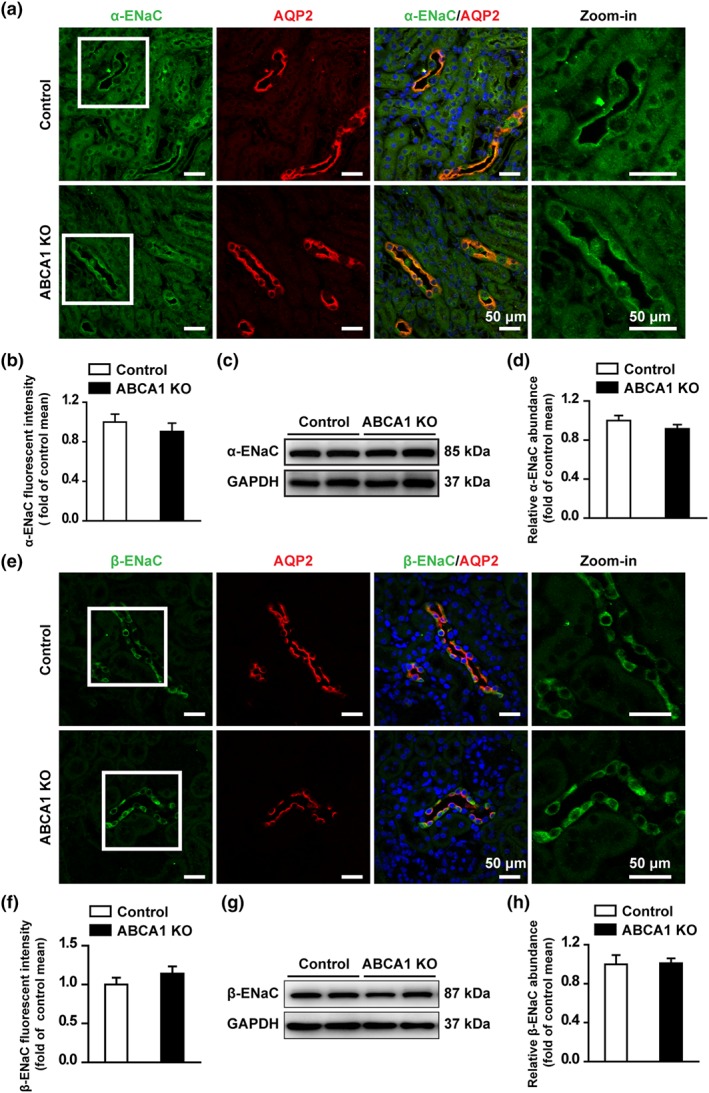
ABCA1 KO does not change the expression of α‐ENaC and β‐ENaC in the CCD of mouse kidney. (a) Confocal microscopy images of α‐ENaC (green) in kidney cortex from control and ABCA1 KO mice. Scale bars: 50 μm. (b) Summary data of α‐ENaC fluorescence intensity in kidney slices from control and ABCA1 KO mice. Each experiment was repeated three times in six mice; 20 images were used for the analysis. (Mann–Whitney test). (c) Western blots of kidney cortex lysates from control and ABCA1 KO mice using antibodies against either α‐ENaC or GAPDH as a loading control. (d) Summary data of Western blots, showing α‐ENaC expression in kidney cortex from control and ABCA1 KO mice (n = 8 in each group). Student's two‐tailed t‐test. (e) Confocal microscopy images of β‐ENaC (green) in kidney cortex from control and ABCA1 KO mice. Scale bars: 50 μm. (f) Summary data of β‐ENaC fluorescence intensity in kidney slices from control and ABCA1 KO mice. Each experiment was repeated three times in six mice; 20 images were used for analysis (Mann–Whitney test). (g) Western blot of kidney cortex lysates from control and ABCA1 KO mice, using antibodies against either β‐ENaC or GAPDH as a loading control. (h) Summary data of Western blots, showing β‐ENaC expression in kidney cortex from control and ABCA1 KO mice (n = 8 in each group). Student's two‐tailed t‐test
