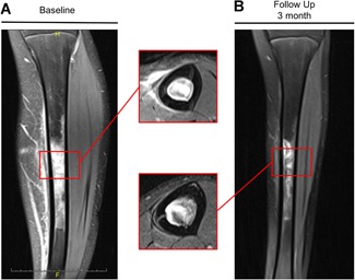
Figure 2.

Coronal T2 MR images of a 14‐year‐old female patient at (A) initial presentation and (B) at follow‐up after 3 months demonstrating bone marrow edema of the tibia diaphysis. The patient was an active handball player and suffered from persistent pain at the tibia. She was first diagnosed with osteomyelitis and treated with antibiotics until transferred to our institution. Laboratory assessment revealed low alkaline phosphatase activity and elevated pyridoxal‐5‐phosphate levels. TNSALP mutation analysis showed a heterozygous mutation (c.746G>T) and the patient was diagnosed with hypophosphatasia.
