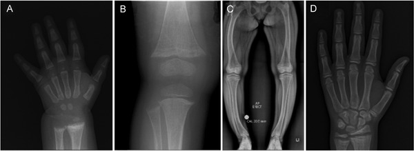
Figure 1.

A now 14‐year‐old female with XLH (PHEX:c.[151C>T];[=] p.[Gln51*]) diagnosed at 7 months of age. (A) Radiograph of left hand and wrist at diagnosis with rachitic changes of distal radius and ulna and lacey appearance of bone. (B) Radiograph of right knee at 18 months old while treated with phosphate and calcitriol. There is fraying and splaying at the metaphyses and early cupping noted at the distal femur as well as the proximal tibia and fibula. (C) At 14 years old, she was managed with phosphate and calcitriol. She had short stature, normal ALP, and mild elevation in PTH. Symptoms included persistent ankle pain and waddling gait. There was also lateral bowing of both femora and tibias with widening of the proximal tibial growth plate. (D) Left hand radiograph at age 14 years showing widening of the proximal radius and ulna growth plates with evidence of rachitic changes.
