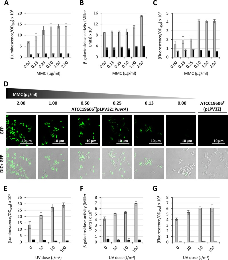
FIG 3.
Regulation of the uvrABC operon upon exposure of A. baumannii to DNA-damaging agents MMC and UV light. Overnight cultures of A. baumannii ATCC 19606T carrying any of the three PuvrA fusions (pLPV1Z::PuvrA or pLPV2Z::PuvrA or pLPV3Z::PuvrA) or the corresponding promoterless vectors were subcultured (1:100 dilution) in LB broth (pLPV1Z::PuvrA or pLPV2Z::PuvrA) or M9-S medium (pLPV3Z::PuvrA) and incubated at 37°C until the culture reached the mid-exponential phase (OD600 of 0.6). Cultures were treated with different MMC concentrations, as indicated, and luminescence (A), β-galactosidase activity (B), and fluorescence emission (C) were measured after 16 h. GFP-producing cells were also visualized using a Leica SP5 confocal laser scanning microscope equipped with a 63× oil immersion objective (D). Representative images of either GFP or GFP and differential interference contrast (DIC)-merged channels are shown. Scale bar, 10 μm. Overnight cultures were also suspended in M9-S medium at an OD600 of 1.0, and 5 ml of each suspension was irradiated with 0, 10, 50, and 100 J/m2 UV light at 300 nm. Luminescence (E), β-galactosidase activity (F), and fluorescence emission (G) were measured after a 2-h rescue at 37°C. Gray and black histograms indicate strains carrying individual PuvrA fusions and the promoterless vectors, respectively. Data are the means ± standard deviations from three independent experiments.

