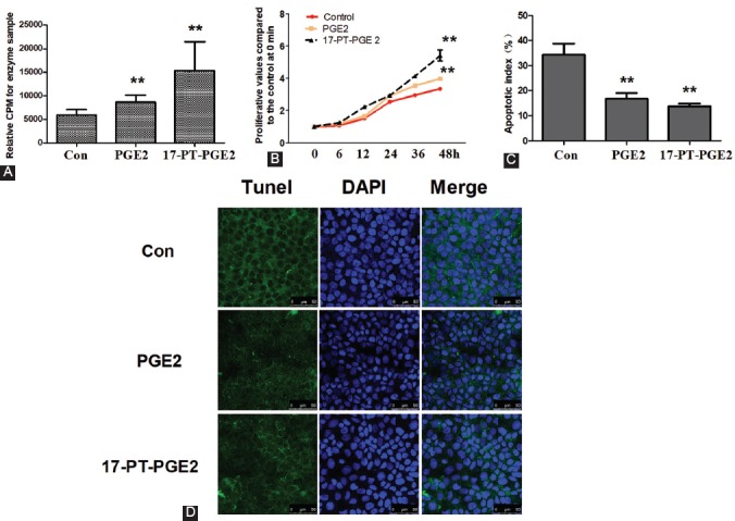
FIGURE 2.

Activation of the EP1 receptor promoted proliferation in osteosarcoma (OS) cells. MG63 cells were cultured with PGE2 (5 µM) or 17-PT-PGE2 (5 µM) for 48 hours. A) PKC activity assay. MG63 cells were treated with 17-PT-PGE2 for 48 hours. Equal amounts of total proteins (30 mg) were added to microcentrifuge tubes and assayed for PKC levels using a PKC activity assay kit. B) Cell viability was tested by MTT assay. C-D) Following the treatment, MG63 cells were fixed and stained with DAPI and TUNEL. The apoptotic nuclei were visualized and photographed using a laser confocal microscope. Each sample was prepared in triplicate and all experiments were repeated three times. All bar/line graphs represent the mean ± SD. **p < 0.01 vs. DMSO-treated control (con). Both PGE2 and 17-PT-PGE2 increased the activity of PKC compared to control cells. Furthermore, the treatment with PGE2 and 17-PT-PGE2 increased the proliferation and decreased the apoptosis of MG63 cells compared to control cells. PGE2: Prostaglandin E2; PKC: Protein kinase C.
